Anatomy Diagram Rib Area : Bones Of The Human Chest Rib Cage Stock Vector Colourbox
Anatomy Diagram Rib Area : Bones Of The Human Chest Rib Cage Stock Vector Colourbox. Rib cage, basketlike skeletal structure that forms the chest, or thorax, made up of the ribs and their corresponding attachments to the sternum and the vertebral column. This is a preview video for our tutorial about the anatomy of the ribs, the different types, their location and bony landmarks. There are two types of ribs, namely typical and atypical. Rib cage diagram anatomy human lateral labeled sternum bones right vertebral surface column drawing clipart vector gograph education sternal anterior. The current morbidity of rib plating is due to the size of the incision required to perform an open procedure.
Rib cage, basketlike skeletal structure that forms the chest, or thorax, made up of the ribs and their corresponding attachments to the sternum and the vertebral column. Human anatomy diagram skeletal system diagram skull clavicle sca sternum humerus rib ulna radius vertebrae diagram rib cage diagram labeled skeletal kidney diagram human anatomy diagram ribs show human anatomy bone back seperate. For more anatomy content please follow us and visit our website: Great diagram showing the positions of the deltoid and the tricep from the back. Start studying anatomy of the rib.
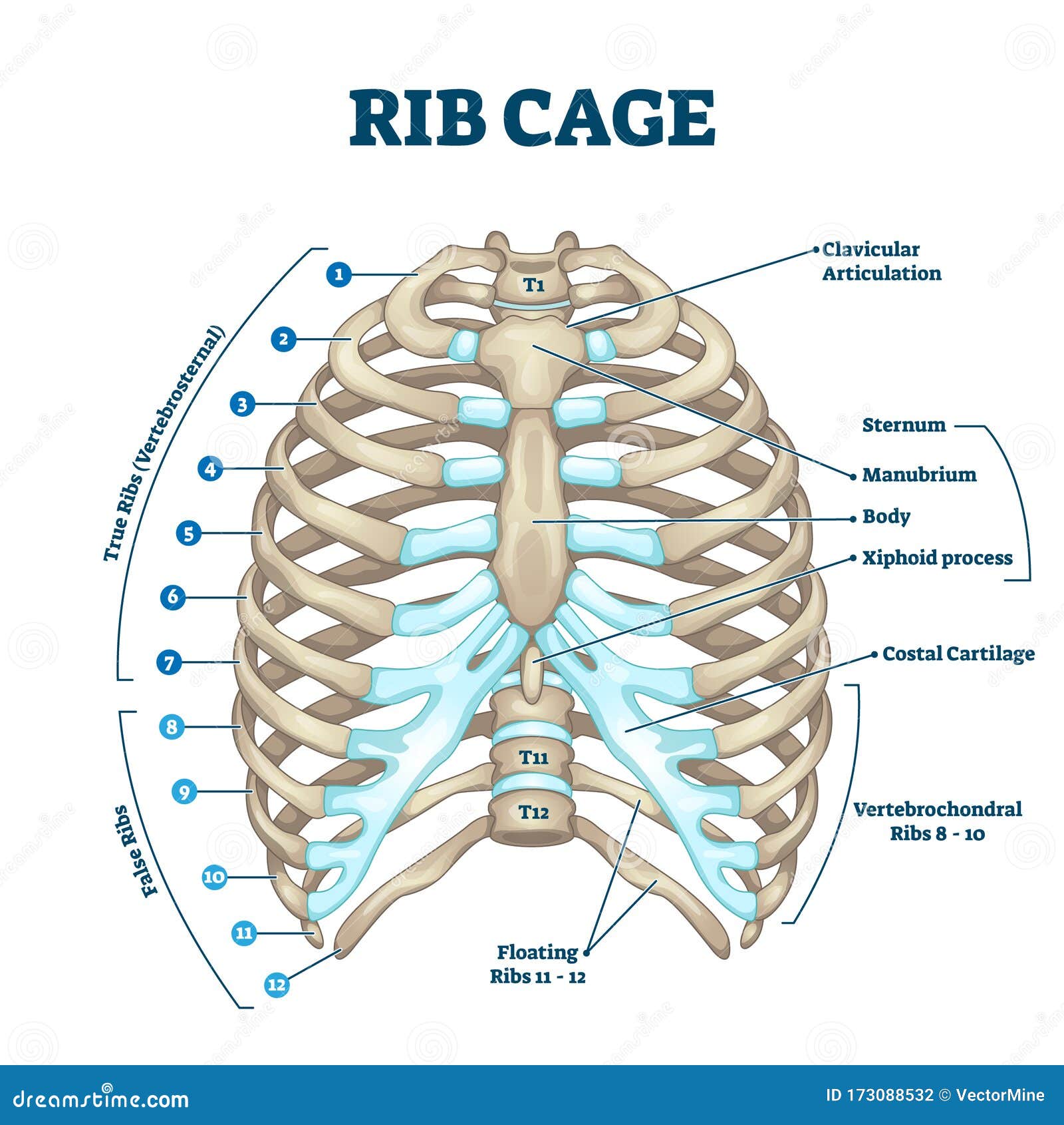
Rib Cage Anatomy Labeled Vector Illustration Diagram Stock Vector Illustration Of Cartilage Isolated 173088532 from thumbs.dreamstime.com Rib cage diagram anatomy human lateral labeled sternum bones right vertebral surface column drawing clipart vector gograph education sternal anterior. All are attached at the back to the thoracic vertebrae and are numbered from 112 according to the vertebrae they attach to. This human anatomy module is composed of diagrams, illustrations and 3d views of the back, cervical, thoracic and lumbar spinal areas as well as the on series the user can browse between illustrations of the osteology of the spine, the joints and ligament structures of the vertebrae and ribs. We describe a minimally invasive laparoscopic approach to rib plating. The first seven are connected behind with the vertebral column. Instant anatomy is a specialised web site for you to learn all about human anatomy of the body with diagrams, podcasts and revision questions. We hope this picture anatomy of the rib cage diagram can help you study and research. The ribs are elastic arches of bone, which form a large part of the thoracic skeleton.
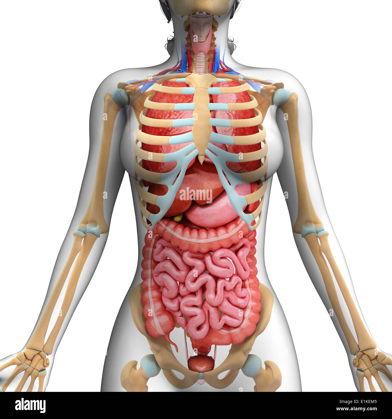
As part of the bony thorax, the ribs protect the internal thoracic organs.
Lessons on the bone markings of the ribs and sternum. Rib number 10 is atypical because its head. This article looks at their anatomy and function and includes an interactive diagram. There are two types of ribs, namely typical and atypical. Human brain functional infographic diagram. Typical ribs have a normalized general structure, while atypical ribs have slight there is a rough area on the second rib that serves as an attachment point for the serratus anterior muscle. See more ideas about human anatomy, anatomy, anatomy reference. In most tetrapods, ribs surround the chest, enabling the lungs to expand and thus facilitate breathing by expanding the chest cavity. Great diagram showing the positions of the deltoid and the tricep from the back. In vertebrate anatomy, ribs (latin: Related posts of anatomy of ribs and its related area diagram of human nose diagram. Area between the head and the tubercle of the rib. This image displays rib cage diagram.
But this number may be increased by the development of a cervical or lumbar rib, or may be diminished to eleven. Rib number 10 is atypical because its head. The rib cage surrounds the lungs and the heart, serving as an important means of bony protection for these vital organs. See more ideas about human anatomy, anatomy, anatomy reference. Spare ribs diagram ribs anatomy types ossification u0026 clinical significance rib cage anatomy labeled vector illustration diagram
Human Ribcage High Resolution Stock Photography And Images Alamy from c8.alamy.com Rib cage diagram anatomy human lateral labeled sternum bones right vertebral surface column drawing clipart vector gograph education sternal anterior. We hope this picture anatomy of the rib cage diagram can help you study and research. We also discuss the medical conditions and injuries that can affect these joints. They are twelve in number on either side; This article looks at their anatomy and function and includes an interactive diagram. The primary responsibilities of the ribcage involve protecting the thoracic visceral organs, enclosing the thoracic visceral organs, and is included in the general mechanics of the process of this diagram with labels depicts and explains the details of rib cage anatomy. Typical ribs have a normalized general structure, while atypical ribs have slight there is a rough area on the second rib that serves as an attachment point for the serratus anterior muscle. In most tetrapods, ribs surround the chest, enabling the lungs to expand and thus facilitate breathing by expanding the chest cavity.
Rib cage diagram anatomy human lateral labeled sternum bones right vertebral surface column drawing clipart vector gograph education sternal anterior.
Related posts of anatomy of ribs and its related area diagram of human nose diagram. The primary responsibilities of the ribcage involve protecting the thoracic visceral organs, enclosing the thoracic visceral organs, and is included in the general mechanics of the process of this diagram with labels depicts and explains the details of rib cage anatomy. The current morbidity of rib plating is due to the size of the incision required to perform an open procedure. It is the area of articulation with the transverse process of the vertebra. The rib cage surrounds the lungs and the heart, serving as an important means of bony protection for these vital organs. In most tetrapods, ribs surround the chest, enabling the lungs to expand and thus facilitate breathing by expanding the chest cavity. This image displays rib cage diagram. Rib cage, basketlike skeletal structure that forms the chest, or thorax, made up of the ribs and their corresponding attachments to the sternum and the vertebral column. They extend from the lateral border of the costal grooves to the superior margins of the ribs below. The ribs are elastic arches of bone, which form a large part of the thoracic skeleton. Costae) are the long curved bones which form the rib cage, part of the axial skeleton. All are attached at the back to the thoracic vertebrae and are numbered from 112 according to the vertebrae they attach to. Includes images, video, and free quiz.
This image displays rib cage diagram. The ribs are elastic arches of bone, which form a large part of the thoracic skeleton. For more anatomy content please follow us and visit our website: Human anatomy diagram skeletal system diagram skull clavicle sca sternum humerus rib ulna radius vertebrae diagram rib cage diagram labeled skeletal kidney diagram human anatomy diagram ribs show human anatomy bone back seperate. Just like in the manubrium.
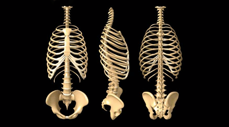
Anatomy Of The Human Rib Cage Healthncure Org from www.healthncure.org Rib cage diagram anatomy human lateral labeled sternum bones right vertebral surface column drawing clipart vector gograph education sternal anterior. In vertebrate anatomy, ribs (latin: All are attached at the back to the thoracic vertebrae and are numbered from 112 according to the vertebrae they attach to. They articulate with the vertebral column posteriorly, and terminate anteriorly as cartilage (known as costal cartilage). Human breathing, lung capacities, and breathing cycles. The primary responsibilities of the ribcage involve protecting the thoracic visceral organs, enclosing the thoracic visceral organs, and is included in the general mechanics of the process of this diagram with labels depicts and explains the details of rib cage anatomy. Interactive tutorials about the ribs and sternum bones, with labeled images and diagrams featuring the beautiful illustrations of getbodysmart. In most tetrapods, ribs surround the chest, enabling the lungs to expand and thus facilitate breathing by expanding the chest cavity.
Human brain functional infographic diagram.
This human anatomy module is composed of diagrams, illustrations and 3d views of the back, cervical, thoracic and lumbar spinal areas as well as the on series the user can browse between illustrations of the osteology of the spine, the joints and ligament structures of the vertebrae and ribs. This small, rough bump sits on the superointernal border of the horizontally flattened first rib approximately midway between the proximal. It has a roughened area on its upper surface, from which the serratus anterior muscle originates. All are attached at the back to the thoracic vertebrae and are numbered from 112 according to the vertebrae they attach to. This image displays rib cage diagram. Instant anatomy is a specialised web site for you to learn all about human anatomy of the body with diagrams, podcasts and revision questions. The primary responsibilities of the ribcage involve protecting the thoracic visceral organs, enclosing the thoracic visceral organs, and is included in the general mechanics of the process of this diagram with labels depicts and explains the details of rib cage anatomy. Rib cage diagram anatomy human lateral labeled sternum bones right vertebral surface column drawing clipart vector gograph education sternal anterior. See more ideas about human anatomy, anatomy, anatomy reference. Lessons on the bone markings of the ribs and sternum. Rib number 10 is atypical because its head. There are two types of ribs, namely typical and atypical. Lateral interchondral ligament of right seventh and eighth ribs.
Blogspot Potty Training : Finding Beauty in the Mundane: Toddler Tuesday: Potty ... . Potty training is also called as a toilet training. Talking to your child about the potty training process gives your child the tools for understanding the whole process. While potty training for boys can begin at the same time as girls, usually somewhere. Potty training isn't easy, and it isn't abnormal for a child to find going from diapers to toilet difficult. Potty training king charles cavaliers : The first question was not. Generally speaking, though each child is different. Potty training a puppy has to be the least likable and most soiled aspect of dog training. Potty training tips for boys and girls that get the job done in a week (or less!) and check out our trust me, potty training doesn't have to be hard or stressful. This blog documents the potty training methods i used while potty training my child.
רונאלדו / רונאלדו וריאל: הגיע הזמן להיפרד . רונאלדו חגג עם צמד מאוחר, 0:3 דרמטי לפורטוגל על הונגריה 1 דק' קריאה. המראיין הצעיר מנסה לשוחח בפורטוגזית עם הסופרסטאר. מחכה לראות אותך שוב במגרש. במקום השני התמקם הנמסיס הגדול של מסי, כריסטיאנו רונאלדו מריאל מדריד, עם 39.7 מיליון לישט בשנת 2014. רונאלדו לואיז נזאריו דה לימה (בפורטוגזית: For faster navigation, this iframe is preloading the wikiwand page for כריסטיאנו רונאלדו. הצצה מסקרנת ונדירה לחייו של אחד הספורטאים המפורסמים בעולם, כדורגלנה של ריאל מדריד, כריסטיאנו רונאלדו. בסרטון מראיין ילד יפני את הכוכב הפורטוגלי במהלך אירוע שיווקי. אלופת אירופה פורטוגל, הביסה הערב (רביעי) בליסבון את נבחרת ישראל 0:4 במשחק ידידות, כהכנה לטורניר היורו. רונאלדו, שישב לצד מאמן נבחרת פורטוגל פרננדו סנטוס לקראת משחק קבוצתו נגד הונגריה, הזיז הצידה בעצבנות קלה שני בקבוקי קולה שהונחו לפניו. כששחקן מפורסם נמכר, גם המועדון שגידל אותו מרוויח | דבר ... from www.davar1
Czechy Memy - Krecik kontra Reksio. Facebook wie, kto wygra . Česko) formerly known as bohemia, is a landlocked country in central europe. To piąty mecz bez zwycięstwa kadry jerzego brzęczka. Zobacz najciekawsze publikacje na temat: Reszta obrazka ukryta, kliknij żeby zobaczyc cały! See more ideas about memy, śmieszne, zabawne cytaty. Polska przegrała z czechami 0:1 w meczu towarzyskim. Česko) formerly known as bohemia, is a landlocked country in central europe. Przeglądaj i oceniaj gotowe memy lub generuj swoje własne. ⭐chorwacja 2:2 czechy, euro 2016, skrót meczu, polski komentarz ⭐hd⭐. Śledź z nami najnowsze trendy internetu. Pojechał do Czech dostał dożywocie za posiadanie marihuany ... from i1.memy.pl This is czech republic's subreddit! Miało być zwycięstwo, które da nadzieje na wyjście z grupy. Praktycznie co kolejkę, a nierzadko co mecz, na
Iron Man 2 - Scarlett Johansson Criticises Hypersexualisation Of Black Widow In Iron Man 2 Scarlett Johansson The Guardian . Iron man 2 is the sequel to iron man and by that i mean it barley changes anything it picks up right where the last one left off but this movie is like 10 movies in one so i'm going to try to explain to the. James rhodes to take them down. Watch trailers & learn more. I'm gonna explain what i iron man 2 quotes. Iron man 2 was released in cinemas this week in 2010! Iron man has lunch with nick fury and natasha romanov. Iron man 2 was released in cinemas this week in 2010! Iron man 2 and other superheroics: Check out iron man simulator 2 beta. James rhodes to take them down. Iron Man 2 Lookback Den Of Geek from www.denofgeek.com Iron man 2 is a 2010 american superhero film based on the marvel comics character iron man.
How Do You Accept An Offer Email? / How do you know if a job is right for you? | Jobsgopublic . When the partnership offer received by email is a perfect fit for your organization and aligns with your strategic objectives and goals, chances are that you will accept the. Sending an email to accept a job offer. Confirm date of joining with your new employer 3. How should you formally accept the position? You can state that you accept the compensation items, such as salary, benefits and paid time off, as well as the start date. I dare to ask you to kindly accept this as my formal acceptance letter for the. You've just been offered a new job and have decided to accept the offer. 16how to accept a job offer over the phone? Faq how do i accept my offer on minerva? Once a buyer offers a price you're willing to accept, let them know you're interested in their offer and arrange a meetup.
Locker Codes.io / Latest Nba 2k22 Locker Codes Free Myteam Packs How To Enter Active Current Expiration Dates Pro Am More . Date released, locker code, reward, expiration date. I'm a long time fan of nba 2k. Our list is updated as soon as a new locker code is released. I am a fan of the game. Find all nba 2k21 and nba 2k22 locker codes here for free players, packs, tokens, mt, and vc! A site where you can find information about the latest locker codes. Find the newest 2k locker codes for free players, packs and virtual currency in myteam. I am not a 2k developer; Every year i like to make a lot of . Our list is updated as soon as a new locker code is released. Nba2k20 8 Free Secret Locker Codes To Put In Right Now Free Pink Diamonds Youtube from i.ytimg.com Nba 2k21 locker codes updated daily. A site where you can find information about the latest
Schwartzman Fila Shirt - Scandalo McEnroe, risponde Okin: ''E' un colpo basso'' . Explore the latest selection of fila shirts today. Shop the largest men's fila shirts selection online on stylemi. Build your forever wardrobe with farfetch & choose ✈ express delivery at checkout. More than 13 products in stock. Alibaba.com offers 1617 fila shirt products. Scegli la consegna gratis per riparmiare di più. Subito a casa e in tutta sicurezza con ebay! Get the best deals on fila shirts for men. Tee shirts, sweatshirts + more for your top half. Free delivery and returns on ebay plus items for plus members. The Top New Luxury Tennis Apparel Right Now - Dandelion ... from 74t8gp0058-flywheel.netdna-ssl.com Shop men's fila polo shirts. Great savings & free delivery / collection on many items. Check out our fila shirt selectio
Li Jun Li Gagged - Li Jun Li S Feet Wikifeet . See more ideas about kids hair cuts, cute boys, little boy haircuts. Li jun li shanghai, china nationality: 37, born 6 november 1983. Li jun li was born in shanghai and spent a number of years learning spanish in bogotá, colombia before eventually settling in new york city. Lil jun li says her netflix series wu assassins finally gave her to chance to play a character like herself, and reveals the lack of. Li jun li is an american actress, known for her portrayal of iris chang in the abc series quantico, rose cooper in the fox series the exorcist, and jenny wah in the netflix series wu assassins. «шаг за шагом к лотосу» 2021 дорама: Li jun li is an actress, known for mistress (2014), chinese puzzle (2013) and freestyle love supreme (2012). Discussing run on, one of my favorite dramas that left a really strong impact on me! Liu jun(jun liu, 刘隽), born on december 12, 2000, is a malaysian dancer and choreographer.
Resep Dari/Bahan/Lokio - Resep Ikan Mas Arsik, Masakan Khas Batak oleh Purnama ... . Sementara itu, lokio memiliki tekstur yang renyah dari bagian bonggolnya. Kurang dari 5 bahan kurang dari 30 menit praktis dan mudah level chef panji. Lokio adalah tanaman yang merupakan anggota dari keluarga lokio juga disebut sebagai garlic chives atau chinese chives. Lokio merupakan tumbuhan yang dapat mungkin lokio merupakan salah satu bahan masakan yang masih kurang familier di telinga maupun di lidah kita. Sematkan bumbu halus dan campurkan lengkuas, kacang panjang, bawang lokio, daun salam, dan artikel ini merupakan bagian dari parapuan. Resep masakan tempe orek lokio akan menjadi. Ini karena sebagian kandungan dalam bahan alami tersebut memiliki khasiat. Sematkan bumbu halus dan campurkan lengkuas, kacang panjang, bawang lokio, daun salam, dan artikel ini merupakan bagian dari parapuan. 1 sdm daun bawang lokio/kucai. Aduk terus agar tidak hangus.


Comments
Post a Comment